|
Researchers from the Argonne National Laboratory near Chicago, USA, along with the Max-Planck-Institut für Kernphysik (MPIK) in Heidelberg and the European X-ray Free Electron Laser (European XFEL) in Hamburg, developed an innovative approach to X-ray spectroscopy, achieving unprecedented detail and resolution.
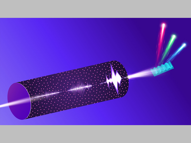
The technique, called stochastic Stimulated X-ray Raman Scattering (s-SXRS), uses intense X-ray pulses to excite electrons within atoms. It was implemented in a pioneering experiment at the European XFEL. The researchers directed an X-ray beam through a small, 5-millimeter high-pressure gas cell designed at MPIK filled with neon as a target gas. The resulting radiation is collected and analysed in a grating spectrometer — a device that separates light into its different wavelengths. As the X-rays pass through the gas, they amplify by nearly a billion-fold the resonantly scattered radiation – so-called Raman signals, a type of X-ray fingerprint that provides information about the excited electronic states of atoms as well as molecules.
The amplified signal provides detailed information about the electronic structure of the atoms on a femtosecond timescale (i. e. a millionth of a billionth of a second). A statistical method, called covariance analysis, links the incoming X-ray pulses with the Raman signals emitted from the atoms. Using this, the scientists create a detailed energy spectrum from many individual snapshots, rather than scanning slowly across different energy levels. What was once considered “noise”, is thus transformed into a valuable resource, allowing extraction of detailed information from complex data.
The large number of photons in each X-ray flash not only boosts the measurement signal but also holds the key to the highest spectral resolution by averaging over many photon impacts on the detector. This large number of photons at random, but fully correlated wavelengths allows to pinpoint the position of the centre of these broad but distinct spectral spikes much more precisely than their width would otherwise indicate. This approach is similar to the super-resolution microscopy technique that won the 2014 Nobel Prize in chemistry.
An essential support for the interpretation of the experimental results were large-scale simulations of the complex interactions of the X-ray pulses while they propagate through the gas. Argonne Leadership Computing Facility (ALCF), provided the necessary computational power for these calculations which closely match the measured data and thus confirming the researchers’ understanding of these processes, paving the way for future investigations. With continued advancements, s-SXRS could become a standard tool in laboratories worldwide, driving innovation across many fields and setting the stage for breakthroughs in chemical analysis and materials science impacting industries like electronics and nanotechnology.
Original publication:
Super-resolution Stimulated X-ray Raman Spectroscopy
Kai Li, Christian Ott, Marcus Agåker, Phay J. Ho, Gilles Doumy, Alexander Magunia, Marc Rebholz, Marc Simon, Tommaso Mazza, Alberto De Fanis, Thomas M. Baumann, Jacobo Montano, Nils Rennhack, Sergey Usenko, Yevheniy Ovcharenko, Kalyani Chordiya, Lan Cheng, Jan-Erik Rubensson, Michael Meyer, Thomas Pfeifer, Mette B. Gaarde and Linda Young
Nature 643, 662 (2025). DOI: 10.1038/s41586-025-09214-5
Weblinks:
Division “Quantum dynamics and control” at MPIK
Group “Excited atoms and molecules in strong fields” at MPIK
„Behind the paper“ blog (Argonne National Laboratory)
Press release (Argonne National Laboratory)